
TSH Levels for Pregnancy & Fertility: Thyroid Health Guide
Couples trying to conceive often spend months tracking ovulation, timing intercourse, and checking pelvic anatomy. Yet an invisible metabolic regulator rests quietly in the neck. Thyroid steers fertility. Cells listen. Every single reproductive tissue possesses nuclear thyroid hormone receptors that dictate cellular energetic velocity. When circulating levels drift even slightly off target, human reproduction stalls. Life pauses. Conception fails. At my consulting chambers in Panchpakhadi, Thane West, I frequently evaluate hopeful couples who feel completely exhausted by unexplained reproductive delays. Their basic pelvic ultrasound scans appear normal. Their semen reports seem acceptable on paper. Despite this reassuring picture, pregnancy does not happen. In many of these cases, the missing link sits directly within endocrine cross-talk, specifically how subtle thyroxine imbalances disrupt cellular signaling inside the ovary, uterus, and testes.
Understanding tsh levels for pregnancy provides couples with actionable clarity. The pituitary gland produces Thyroid-Stimulating Hormone (TSH) to tell your thyroid how much thyroxine (T4) and triiodothyronine (T3) to manufacture. When circulating thyroid hormones drop, the pituitary secretes more TSH to urge the thyroid onward. When thyroid hormone levels rise, TSH drops. What constitutes a standard lab range for an ordinary adult, however, does not support early embryonic life. Modern reproductive endocrinology establishes clear guidelines: healthy conception demands far tighter metabolic control than standard wellness screenings require.
Endocrine Cross-Talk: The Hypothalamic-Pituitary-Ovarian Axis
Reproductive biology does not function in isolation. Glands talk constantly. At the base of the brain, the hypothalamus produces Thyrotropin-Releasing Hormone (TRH). This neurohormone stimulates the anterior pituitary gland to release TSH. When someone develops subclinical or overt hypothyroidism, the hypothalamus secretes excess TRH to stimulate the struggling thyroid.
Excess TRH triggers lactotroph cells within the pituitary. This stimulation causes mild hyperprolactinemia. Elevated prolactin disrupts the pulsatile release of Gonadotropin-Releasing Hormone (GnRH). Without clean GnRH pulses, the pituitary fails to release appropriate ratios of Luteinizing Hormone (LH) and Follicle-Stimulating Hormone (FSH). Follicular recruitment slows down. Anovulation or irregular menstrual cycles follow. Couples struggling with metabolic delays also benefit from managing PCOS and insulin resistance alongside thyroid screening, as these conditions frequently overlap.
Beyond pituitary feedback, thyroid hormones directly nourish growing follicles. Cells need thyroxine. Ovarian granulosa cells feature dense T3 nuclear receptor populations. Here, triiodothyronine cooperates with FSH to stimulate healthy steroidogenesis, encouraging granulosa cell survival and strong aromatase activity. When thyroid hormone concentrations drop below physiological targets, developing oocytes suffer energetic compromise.
Luteal Phase Deficiency and Endometrial Receptivity
Conception requires more than simple ovulation. Implantation demands precision. Timing rules biology. Once a mature follicle successfully expels an oocyte during the mid-cycle luteinizing hormone surge, remaining granulosa and theca cells rapidly reorganize into the highly vascularized corpus luteum to initiate continuous steroidogenesis. Vessels grow fast. The corpus luteum must pump out substantial progesterone for ten to fourteen days to prepare the uterine lining for an arriving blastocyst.
Thyroid hormones stimulate human luteal cells directly. In subclinical hypothyroidism, granulosa cells cannot sustain prolonged progesterone production. The luteal phase collapses prematurely to nine or ten days. Patients observe this failure as persistent brown premenstrual spotting or abbreviated menstrual cycles. Even if fertilization occurs inside the fallopian tube, the blastocyst reaches a hostile uterine environment unable to support apposition.
Decidualization requires adequate intracellular thyroxine. Stromal cells within the endometrium differentiate into specialized decidual cells under the coordinated influence of progesterone and T3. Thyroid hormone deficiency weakens local vascular remodeling and blunts angiogenic factors. Without strong vascular remodeling, the blastocyst cannot burrow into the maternal decidua, culminating in early implantation failure or silent chemical loss.
What Is the Normal TSH Level in Pregnancy and Preconception?
Evaluating clinical tsh levels for pregnancy requires departing from routine adult reference boundaries. Standard laboratory reference ranges on general diagnostic reports usually list TSH between 0.45 and 4.5 or 5.0 mIU/L. While a reading of 4.2 mIU/L may satisfy standard medical screening for a retired sedentary individual, it represents an obstacle for pregnancy.
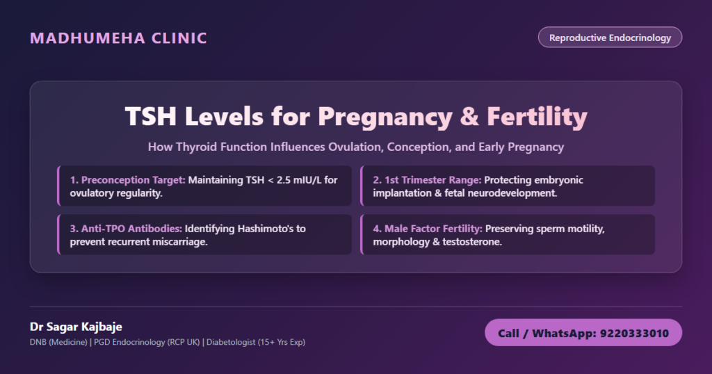
The American Thyroid Association (ATA) and international endocrine consensus state that the preconception TSH target must stay between 0.5 and 2.5 mIU/L. Maintaining a tight normal tsh level in pregnancy ensures that ovarian tissues receive sufficient metabolic stimulation. This threshold provides an protective physiological safety buffer for embryonic development.
| Reproductive Stage / Clinical Scenario | Target TSH & Endocrine Reference | Underlying Pathophysiology | Clinical Action Protocol |
|---|---|---|---|
| Preconception Planning Window | 0.5 to 2.5 mIU/L | Subclinical hypothyroidism blunts granulosa steroidogenesis and shortens the luteal phase. | Screen TSH and anti-TPO antibodies; initiate low-dose levothyroxine if TSH > 2.5 mIU/L. |
| First Trimester Gestation | 0.1 to 2.5 mIU/L (or population trimester range) | Embryo depends entirely on transplacental maternal free T4 for neural development. | Recheck TSH every 4 weeks; increase levothyroxine dosage by 25-30% upon positive pregnancy test. |
| Luteal Phase Deficiency & Recurrent Loss | 1.0 to 2.0 mIU/L; Anti-TPO < 35 IU/mL | Thyroid autoantibodies trigger local decidual micro-inflammation and compromise trophoblast invasion. | Evaluate complete thyroid antibody profile, luteal progesterone, and maternal metabolic markers. |
| Male Subfertility Workup | 0.8 to 2.5 mIU/L; Free T4 within normal limits | Hypothyroidism reduces SHBG, lowers free testosterone, and induces epididymal oxidative stress. | Screen thyroid function in men with unexplained asthenoteratozoospermia; correct before ART. |
Clinical Case: Resolving Secondary Infertility in Panchpakhadi, Thane West
Mrs Ananya Kulkarni (name changed), a 31-year-old corporate marketing manager living in Panchpakhadi, Thane West, visited my clinic after experiencing secondary subfertility for over two years. Her first pregnancy four years earlier had occurred spontaneously without complications. When she and her husband attempted to conceive their second child, nothing happened.
Her pelvic sonography showed normal ovaries with adequate antral follicle counts. Her husband had a normal semen analysis. Ananya felt persistent midday fatigue and noticeable cold intolerance. She also reported that her menstrual cycle had shortened to 24 days with three days of brown premenstrual spotting.
When I evaluated her endocrine profile, her serum TSH measured 4.8 mIU/L. While her previous primary care report marked this as normal, it exceeded the recommended preconception threshold. Her anti-TPO antibodies were elevated at 280 IU/mL. Elevated TRH had generated mild subclinical hyperprolactinemia, blunting her corpus luteum progesterone production and shortening her luteal phase to nine days.
I initiated therapy with levothyroxine 50 mcg taken thirty minutes before breakfast on an empty stomach. Within eight weeks, her TSH settled at 1.8 mIU/L. Her luteal phase lengthened to thirteen days, and her premenstrual spotting stopped. Four months after reaching her target TSH, she conceived spontaneously. We maintained close endocrine supervision throughout her pregnancy, and she delivered a vibrant, healthy baby boy.
The First Trimester: Managing TSH Level in Pregnancy 1st Trimester
Early gestation represents an intense biological stress test for the maternal thyroid gland. During the initial ten to twelve weeks of gestation, the embryonic thyroid has not formed. The embryo depends on maternal transplacental free T4 passing through the placenta.
This maternal thyroxine fuels embryonic neurogenesis, cerebral cortex assembly, and neural tube formation. Human chorionic gonadotropin (hCG), produced by the developing syncytiotrophoblast, shares structural homology with pituitary TSH. In early pregnancy, surging hCG molecules bind directly to maternal TSH receptors on the thyroid gland, stimulating transient increases in thyroid hormone production.
In women with normal glandular reserve, this physiological stimulation suppresses serum TSH, creating the characteristic lower reference range seen in early gestation. Maintaining a stable tsh level in pregnancy 1st trimester below 2.5 mIU/L ensures that the fetus receives continuous hormonal support.
A neglected high tsh level in pregnancy brings documented maternal and fetal hazards:
– Spontaneous first-trimester miscarriage and silent embryonic demise
– Subchorionic hemorrhage and defective trophoblast invasion
– Increased risk of gestational hypertension and preeclampsia
– Neurodevelopmental deficits and lower child intellectual quotients
– Preterm labor and placental abruption
Mothers managing endocrine complications must understand thyroid disorders during pregnancy to prevent sudden hormonal drops that endanger gestation.
Autoimmune Thyroiditis: Hashimoto’s and Anti-TPO Antibodies
Many women trying to conceive show normal TSH readings but carry elevated thyroid autoantibodies. The most common culprit is thyroid peroxidase antibody (anti-TPO), followed by anti-thyroglobulin (anti-Tg). These markers identify Hashimoto’s autoimmune thyroiditis, an autoimmune condition where immune cells target thyroid tissue.
Clinical trials demonstrate that euthyroid women with elevated anti-TPO antibodies carry a two- to three-fold higher risk of first-trimester miscarriage compared to antibody-negative peers. This vulnerability occurs through two primary mechanisms:
1. Limited Thyroid Reserve: When hCG surges in early pregnancy, an inflamed thyroid cannot increase thyroxine production adequately. Mild gestational hypothyroidism develops silently.
2. Endometrial Immune Dysregulation: Elevated autoantibodies reflect broader systemic immune imbalance. Cytotoxic natural killer (NK) cells and inflammatory cytokines invade the endometrium, disrupting embryonic tolerance and leading to micro-thrombosis at the maternal-fetal interface.
For women with positive anti-TPO antibodies preparing for conception, we keep TSH strictly between 1.0 and 2.0 mIU/L. Low-dose levothyroxine shields the early gestational sac against sudden hypothyroxinemia.
Male Factor Subfertility: Thyroid and Fertility in Men
Fertility evaluations often focus exclusively on female biology. This practice overlooks half the equation. Men possess dense populations of thyroid hormone receptors within their testicular architecture. Sertoli cells and Leydig cells require balanced thyroxine signaling to regulate spermatogenesis and testosterone synthesis.
Understanding the dynamic of thyroid and fertility in men uncovers hidden drivers of abnormal semen analyses. When a man develops primary hypothyroidism, several reproductive pathways decline:
– Suppressed SHBG Synthesis: Low thyroid function diminishes hepatic Sex Hormone-Binding Globulin (SHBG) output, lowering total circulating testosterone and altering the testosterone-to-estrogen balance.
– Leydig Cell Inhibition: Blunted thyroxine signaling impairs Leydig cell steroidogenesis, leading to reduced intratesticular testosterone levels necessary for sperm production.
– Epididymal Oxidative Stress: Hypothyroidism elevates reactive oxygen species (ROS) in the male tract, accelerating sperm membrane lipid peroxidation and triggering sperm DNA fragmentation.
– Erectile and Ejaculatory Disturbances: Elevated prolactin and thyroid deficiency cause decreased libido, erectile dysfunction, and delayed ejaculation.
Clinical Case: Correcting Severe Asthenoteratozoospermia in Kalyan
Mr Siddharth Patil (name changed), a 34-year-old software engineer from Kalyan, presented to our center alongside his wife, Pooja (name changed). The couple had been attempting to conceive for three years without success. Pooja’s gynecological workup, ovulation tracking, and tubal patency tests appeared flawless.
Siddharth’s semen analysis showed significant abnormalities. His sperm concentration stood at 18 million/mL, but progressive motility fell to 22%, with normal forms registering at just 2% on Kruger strict criteria (asthenoteratozoospermia). The couple had been advised to proceed immediately with intracytoplasmic sperm injection (ICSI).
Siddharth felt chronic fatigue, muscle lethargy, and experienced modest unexplained weight gain. Because thyroid function is frequently overlooked in male subfertility evaluations, I ordered a detailed endocrine panel. Siddharth had unsuspected overt primary hypothyroidism with a TSH of 14.2 mIU/L and low free T4.
I started Siddharth on levothyroxine 75 mcg daily. His metabolic status normalized steadily. Within ten weeks, his serum TSH settled at 1.9 mIU/L. Because human spermatogenesis requires approximately 74 to 90 days to generate a new cohort of mature spermatozoa, we scheduled a repeat semen analysis three months later.
His repeat semen profile showed remarkable improvement. Sperm concentration reached 46 million/mL, progressive motility doubled to 48%, and normal morphology improved to 6%. Four months later, Pooja conceived naturally, avoiding expensive and invasive assisted reproductive procedures.
Metabolic Synergies: Thyroid, Lipids, and Insulin Resistance
Thyroid function interlocks closely with glycemic and lipid pathways. An underactive thyroid impairs hepatic low-density lipoprotein (LDL) receptor expression, raising circulating cholesterol and triglycerides. Elevated lipids compound subclinical inflammation inside delicate reproductive tissues.
Thyroid hormone deficiency blunts insulin-mediated glucose disposal in peripheral skeletal muscle. This promotes compensatory hyperinsulinemia. Elevated insulin drives ovarian theca cells to overproduce androgens, creating ovulatory arrest. Addressing the broader metabolic effects of thyroid dysfunction on blood sugar and lipids provides couples with durable reproductive recovery.
Clinical Protocol: When and How to Test
Couples experiencing reproductive delays should not delay endocrine testing. Screening should take place early in the investigative cycle.
Tracking maternal tsh levels for pregnancy demands meticulous laboratory assessment. Testing parameters must include:
– Serum TSH: Measured in the morning under fasting conditions.
– Free T4 and Free T3: Assesses bioavailable circulating hormones rather than total levels bound to transport proteins.
– Anti-TPO and Anti-Thyroglobulin Antibodies: Identifies underlying Hashimoto’s thyroiditis before biochemical failure occurs.
– Early Pregnancy Confirmation Protocol: Women with known thyroid dysfunction should test their serum TSH immediately upon a missed menstrual period. Levothyroxine requirements often rise by 25 to 30 percent during the early weeks of gestation.
Frequently Asked Questions
Can thyroid problems cause infertility in women and men?
Yes. Thyroid imbalances alter reproductive capability in both sexes. In women, an underactive thyroid increases TRH secretion, causing elevated prolactin that disrupts GnRH pulsatility, halts ovulation, and shortens the luteal phase. In men, hypothyroidism lowers testosterone synthesis, increases testicular oxidative stress, and produces asthenoteratozoospermia (low sperm count, poor motility, and abnormal morphology).
What are the normal TSH levels for pregnancy and preconception?
The American Thyroid Association recommends keeping serum TSH between 0.5 and 2.5 mIU/L during the preconception period. Once pregnant, the target tsh levels for pregnancy must stay within trimester-specific reference ranges, maintaining TSH below 2.5 mIU/L throughout the first trimester to support embryonic organogenesis.
How does hypothyroidism affect ovulation and the luteal phase?
Hypothyroidism impairs follicular maturation by reducing direct T3 synergy with FSH on ovarian granulosa cells. Elevated TRH triggers hyperprolactinemia, which suppresses LH surges. Following ovulation, inadequate thyroid hormone signaling prevents the corpus luteum from synthesizing sufficient progesterone, causing luteal phase deficiency and early implantation failure.
Why is TSH kept below 2.5 mIU/L in the first trimester of pregnancy?
During the first trimester, the developing fetus has no functioning thyroid gland and relies on maternal thyroxine crossing the placenta for brain and spinal cord development. Keeping TSH below 2.5 mIU/L ensures maternal thyroxine reserves meet fetal neurological demands while minimizing risks of miscarriage, preeclampsia, and placental abruption.
Can elevated thyroid antibodies (Anti-TPO) cause recurrent miscarriages?
Yes. Women with elevated anti-TPO antibodies face a two- to three-fold higher risk of miscarriage even when their TSH appears normal. These antibodies signal underlying autoimmunity that triggers decidual micro-inflammation and impairs trophoblast invasion. They also restrict the thyroid gland from expanding thyroxine production when stimulated by hCG in early gestation.
Medical Review and Clinical Guidance at Madhumeha Clinic
This clinical monograph is authored by Dr Sagar Kajbaje, DNB (General Medicine), Post Graduate Diploma in Diabetes and Endocrinology, Royal College of Physicians (UK), with over 15 years of dedicated clinical experience in reproductive endocrinology, diabetes care, and thyroid medicine.
Couples navigating delayed conception, recurrent pregnancy loss, or complex metabolic conditions can access personalized care locally or through an online endocrine and thyroid tele-consultation across Maharashtra and India.
To schedule an appointment in Thane West, find Madhumeha Clinic on Google Maps. Visit our official portal at Madhumeha Clinic.
Take charge of your reproductive health. Connect directly with our clinical desk: Call: 9220333010 or WhatsApp: 9220333010.
[…] especially when you have diabetes. Water retention, hormones (including thyroid function; read our clinical guide to TSH levels for pregnancy and fertility), and even the time of day can affect what you see on the scale. Instead of relying solely on […]