
Diabetic Bone Disease Prevention: Protect Fragile Bones & Hips
Diabetes doesn’t just affect blood vessels; proactive diabetic bone disease prevention is essential to safeguard bone architecture, prevent micro-fractures, and protect aging joints.
Bones weaken silently. Falls shatter hips. According to national epidemiological estimates, millions of older adults live with brittle skeletons without noticing physical pain until a sudden fracture destroys their mobility. Understanding the medical osteoporosis definition gives individuals with diabetes a clear framework to protect their skeletal health before a minor trip triggers an emergency hospital admission. Density fades slowly. At Madhumeha Clinic, our clinical team frequently sees patients who manage blood sugar diligently while completely overlooking skeletal fragility. Chronic hyperglycemia quietly degrades bone microarchitecture over time. This clinical guide explains how to identify early bone thinning, choose targeted therapies, and protect your independence.
Osteoporosis vs Osteopenia: Key Differences in T-Scores and Fracture Risks
Understanding T-Scores on Bone Densitometry
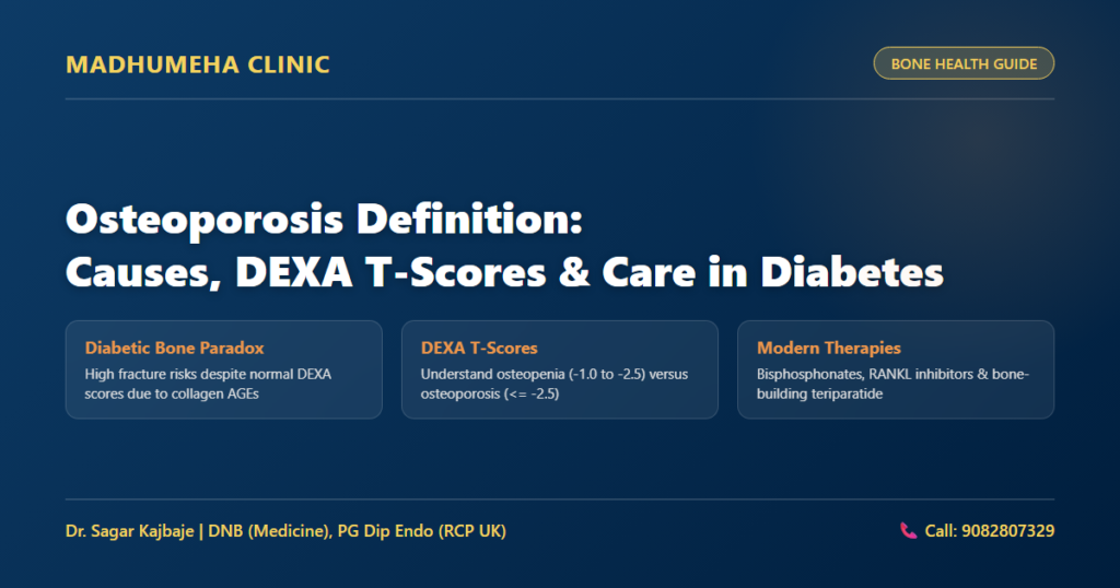
Weak bones break. The World Health Organization established the recognized clinical osteoporosis definition based on dual-energy X-ray absorptiometry measurements of the hip and lumbar spine. A DEXA scanner compares your bone mineral density against the peak bone mass of a healthy thirty-year-old adult. Statisticians express this comparative measurement as a T-score. Under this standard osteoporosis definition, any T-score of -2.5 or lower confirms significant skeletal demineralization. Conversely, clinicians diagnose osteopenia when a patient shows a T-score between -1.0 and -2.5.
Understanding osteoporosis vs osteopenia helps patients recognize why early bone thinning requires proactive intervention rather than passive observation. Many people assume that osteopenia carries little danger. That assumption is dangerous. In clinical practice, more total fractures occur in patients with osteopenia simply because far more people sit in this intermediate score range.
Mrs. Mangala Joshi (name changed) from Thane, a 61-year-old woman living with type 2 diabetes for 14 years, experienced this exact paradox. A trivial trip over a carpet edge fractured her femoral neck. Her DEXA scan revealed a spine T-score of -2.1, which technically indicated osteopenia rather than osteoporosis. Despite this moderate score, advanced glycation end-products had crosslinked her bone collagen, leaving her skeleton remarkably brittle. Dr. Sagar Kajbaje initiated denosumab therapy alongside calcium carbonate and active calcitriol while stabilizing her daily blood glucose levels. This targeted strategy helped her regain independent walking without recurring fractures. Patients with diabetes must track their skeletal markers alongside understanding your HbA1c test results to protect against microvascular and structural bone deterioration.
Factors Causing Osteoporosis in Diabetic and Aging Patients
Cellular Mechanisms and the Diabetic Bone Paradox
Sugar damages bone. Clinicians classify the factors causing osteoporosis into modifiable lifestyle elements and non-modifiable metabolic derangements. Age-related estrogen depletion accelerates osteoclast activity in postmenopausal women. However, longstanding diabetes introduces a destructive metabolic environment that fundamentally alters skeletal biology. Chronic hyperglycemia triggers non-enzymatic glycation of Type 1 collagen, accumulating advanced glycation end-products inside the bone matrix. These molecular crosslinks stiffen collagen fibrils and eliminate the natural elasticity that absorbs mechanical impact. As a result, the skeleton shatters like dry porcelain during low-impact stumbles.
Collagen fractures inside. Beyond matrix damage, high circulating glucose levels suppress osteoblast function, impairing new bone formation and mineral deposition. Diabetic bone disease complicates the classic osteoporosis definition because patients often experience fragility fractures despite having normal bone mineral density scores. Microvascular damage also restricts blood supply to the marrow cavity, depriving osteocytes of needed oxygen and nutrients. At our clinic, we emphasize correcting vitamin D deficiency early because inadequate cholecalciferol compounds insulin resistance and accelerates bone loss.
Mr. Suresh Kadam (name changed) from Kalyan, a 68-year-old gentleman with longstanding type 2 diabetes, sought treatment for chronic, dull lower back discomfort. Spine radiographs revealed an asymptomatic wedge compression fracture in his second lumbar vertebra. His formal bone density test confirmed a spine T-score of -2.8. Through annual zoledronic acid infusions, progressive resistance band training, and home fall-proofing, his back pain settled and his bone density stabilized. Early screening saves mobility.
Bone Density Test Cost and Diagnostic Evaluations in India
Diagnostic Technologies and the Clinical Bone Diagnostic Matrix
Timely testing prevents permanent disability. Patients often ask about the bone density test cost across diagnostic centers in Maharashtra and major Indian cities. A dual-energy X-ray absorptiometry scan of the spine and bilateral hips generally ranges between Rs. 1,500 and Rs. 3,500. Peripheral ultrasound screenings at community health camps offer quick estimates, but they lack the diagnostic precision of axial DEXA scans. Clinicians rely on this diagnostic osteoporosis definition when interpreting diagnostic T-scores alongside clinical risk factors. When evaluating diabetic individuals, doctors frequently calculate a Trabecular Bone Score to evaluate bone microarchitecture directly. Combining DEXA with TBS provides a far clearer picture of true fracture probability.
To help patients understand their diagnostic categories, our clinical team uses this clinical bone diagnostic matrix:
| Bone Health Category | DEXA T-Score Range | Fracture Risk in Diabetes | Clinical Treatment Protocol |
|---|---|---|---|
| Normal Bone Density | -1.0 or higher | Moderate due to diabetic collagen glycation | Lifestyle changes, daily calcium intake, and glycemic control |
| Osteopenia (Low Bone Mass) | Between -1.0 and -2.5 | High due to impaired microarchitecture | Vitamin D repletion, fall prevention, and targeted pharmacotherapy |
| Osteoporosis | -2.5 or lower | Very high with severe risk of hip fractures | Bisphosphonates, RANKL inhibitors, and fall-proofing |
| Severe / Established Osteoporosis | -2.5 or lower with fragility fracture | Extreme risk of recurrent vertebral and femoral collapse | Teriparatide anabolic therapy, antiresorptives, and rehab |
Diagnostic scores guide therapy. If your scan indicates bone loss, your physician will prescribe targeted pharmaceutical therapies to rebuild structural strength.
Choosing the Right Osteoporosis Medicine for Diabetic Patients
Antiresorptive Versus Anabolic Bone Therapies
Medications restore mineral balance. Selecting an appropriate osteoporosis medicine requires careful evaluation of renal function and historical fracture incidence. Antiresorptive agents remain the most common medical class for preserving bone structure. Oral bisphosphonates like alendronate bind to hydroxyapatite crystals and slow osteoclast-mediated bone breakdown. For patients with upper gastrointestinal reflux, doctors often prescribe annual intravenous zoledronic acid infusions. However, clinicians must check serum creatinine and estimated glomerular filtration rate prior to infusion because impaired kidneys cannot clear zoledronic acid safely.
Cells remodel bone tissue. Denosumab represents another powerful antiresorptive option. Given as a subcutaneous injection every six months, denosumab neutralizes RANK ligand, halting osteoclast survival. Unlike bisphosphonates, denosumab does not rely on renal excretion, making it suitable for diabetic individuals with mild to moderate chronic kidney disease. For patients suffering from severe osteoporosis with vertebral fractures, teriparatide provides an anabolic mechanism. This recombinant human parathyroid hormone stimulates osteoblasts directly, driving rapid formation of new trabecular bone. Every pharmaceutical regimen also requires adequate nutritional support. Adults require 1,000 to 1,200 milligrams of elemental calcium daily from dietary sources or supplements. Clinicians also prescribe 60,000 international units of cholecalciferol weekly for eight weeks to correct underlying deficiency before moving to monthly maintenance.
Modern Osteoporosis Treatments: From Bisphosphonates to Lifestyle Changes
Exercise Prescriptions, Fall Prevention, and Glycemic Control
Bones demand mechanical stress. Evidence-based osteoporosis treatments combine targeted pharmacology with structured physical rehabilitation. Sedentary living speeds up calcium loss from weight-bearing bones. When muscles pull against skeletal attachment points, osteocytes detect mechanical strain and trigger localized bone remodeling. Engaging in structured strength training routines builds muscular stability while stimulating trabecular bone mineral accumulation. Brisk walking, light resistance bands, and bodyweight squats provide safe skeletal loading without excessive impact.
Falls shatter hips. Preventing domestic slips forms a key pillar of home safety. Diabetic peripheral neuropathy reduces foot sensation, while diabetic retinopathy compromises depth perception in dim lighting. Families should remove loose scatter rugs, install sturdy grab bars beside toilets, and place bright nightlights in hallways. Correcting glycemic instability also prevents sudden episodes of hypoglycemic dizziness that precipitate catastrophic domestic falls. Protecting bone requires consistent, daily attention across nutrition, movement, and medical oversight.
Dedicated Bone Health Care at Madhumeha Clinic
Evidence-Based Diabetes and Osteoporosis Management
Strong bones preserve your freedom of movement. At Madhumeha Clinic, Dr. Sagar Kajbaje provides clinical guidance for diabetic patients dealing with osteopenia and bone loss. Dr. Kajbaje holds a DNB in General Medicine and a Post Graduate Diploma in Diabetes and Endocrinology from the Royal College of Physicians (UK). He brings over 15 years of specialized clinical experience to complex bone and metabolic disorders. Our consultation fee is Rs. 300 for both first consultation and follow-up visits. To schedule an evaluation, call Call: 9082807329 or book an online diabetes tele-consultation.
Frequently Asked Questions
Can osteoporosis be reversed or cured?
Medical therapies cannot completely cure established osteoporosis, but modern interventions can substantially rebuild bone mineral density and restore structural integrity. Anabolic medications like teriparatide actively stimulate new bone matrix formation, while antiresorptive drugs prevent further osteoclastic breakdown. Combining prescription pharmacotherapy with progressive resistance exercises and adequate calcium restores significant skeletal strength.
How is osteoporosis diagnosed in diabetic patients?
Physicians diagnose osteoporosis primarily through a dual-energy X-ray absorptiometry scan of the spine and hips. Under the standard osteoporosis definition, doctors confirm bone loss when a DEXA scan shows a T-score of -2.5 or below. However, diabetes creates brittle collagen without always lowering mineral density. Clinicians therefore evaluate Trabecular Bone Scores and prior fracture history to gauge actual skeletal vulnerability.
What are the warning signs and symptoms of osteoporosis?
Bone loss produces zero early warning symptoms. Most individuals notice no discomfort until a sudden low-impact fall causes a painful fracture in the hip, wrist, or spine. Advanced bone thinning may cause gradual height loss, progressive stooping of the upper back, and persistent postural back pain from silent vertebral compression.
Why does diabetes increase the risk of osteoporosis and bone fractures?
High blood glucose levels damage bones through multiple biological pathways. Chronic hyperglycemia causes advanced glycation end-products to crosslink Type 1 collagen, making the skeletal matrix stiff and brittle. Additionally, diabetes impairs osteoblast activity, reduces bone blood flow, and causes peripheral neuropathy that increases fall frequency.
How often should a DEXA bone density scan be performed?
Clinicians generally recommend a repeat DEXA bone density scan every one to two years for patients receiving active osteoporosis treatments. This interval allows doctors to assess therapeutic response, confirm bone density stability, and modify medical regimens when necessary. Patients with stable bone mass and well-controlled diabetes may test every two to three years depending on clinical risk factors.
[…] bone disease, DEXA T-scores, and fracture prevention, read our comprehensive medical guide on osteoporosis definition covering bone density testing and modern […]