
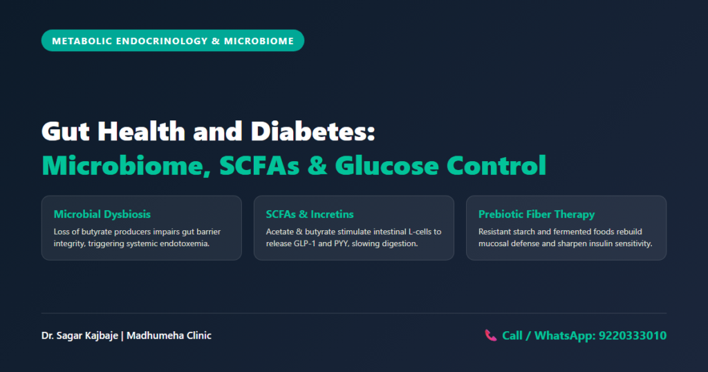
Gut Bacteria and Diabetes: The Microbiome Blood Sugar Link
Every week at Madhumeha Clinic in Panchpakhadi, Thane West, patients sit across from me looking thoroughly defeated by their glucometer numbers. Many follow strict calorie counts. They take their prescribed medications on schedule. Yet, their fasting blood sugars stay persistently high, and their afternoon numbers swing wildly. For decades, traditional clinical medicine treated Type 2 diabetes solely as an endocrine defect of pancreatic beta-cells and skeletal muscle insulin resistance. Modern research reveals another reality. Bugs control sugar. Microbes matter. Trillions of microorganisms residing inside your gastrointestinal tract actively dictate how your body processes carbohydrates, triggers systemic inflammation, and regulates insulin sensitivity. Understanding the clinical link between gut bacteria and diabetes is transforming how we reverse metabolic dysfunction.
Microbes dictate metabolism. Your colon houses roughly thirty-eight trillion bacterial cells representing more than one thousand distinct species. When this inner ecosystem functions in harmony, these microorganisms act as an active metabolic organ. They digest complex plant polysaccharides, synthesize micronutrients, and produce biochemical signaling molecules that communicate directly with your liver, pancreas, and brain. When poor lifestyle habits destroy this ecosystem, metabolic chaos ensues. Examining your inner flora provides clinical answers that routine calorie counting cannot explain. Stool reveals secrets. Tests guide care. Fibers feed flora.
The Gut Microbiome and Diabetes: What Science Tells Us
The human digestive tract contains two dominant bacterial phyla known as Firmicutes and Bacteroidetes. Healthy individuals maintain a balanced ratio between these two groups, alongside thriving populations of protective commensal organisms such as Faecalibacterium prausnitzii, Roseburia, and Akkermansia muciniphila. In patients with metabolic disease, this delicate architectural balance collapses. This clinical state of microbial disruption is termed dysbiosis.
Good bugs vanish. Bad bugs thrive. Ecology collapses.
Decades of clinical stool sequencing confirm that patients with Type 2 diabetes exhibit a characteristic microbial signature. Studies on gut bacteria and type 2 diabetes show that protective species decline sharply. Harmful Gram-negative opportunistic pathogens expand rapidly. Research examining the gut microbiome and diabetes indicates that this compositional collapse directly undermines glucose homeostasis and physiological resilience. Research into gut bacteria and diabetes reveals that losing beneficial taxa damages the gut lining, impairs carbohydrate fermentation, and disrupts normal bile acid signaling.
Species diversity drops. Metabolic health requires diversity. When your internal bacterial garden loses its richness, your physiological resilience declines. Restoring microbial species composition forms the foundation of modern diabetes management.
+-----------------------------------------------------------------------------------+
|                        THE GUT-METABOLIC CASCADE                                  |
|                                                                                   |
|   Poor Diet / Dysbiosis           Intestinal Barrier Breakdown (Leaky Gut)        |
|   (Depleted Akkermansia)   --->   (Elevated Zonulin, Damaged Tight Junctions)     |
|                                                     |                             |
|                                                     v                             |
|   Insulin Resistance & High Blood Sugar <--- Systemic Low-Grade Inflammation      |
|   (TLR4 Activation on Liver/Muscle)          (Bacterial LPS Translocation)        |
+-----------------------------------------------------------------------------------+
Does Gut Bacteria Affect Blood Sugar Levels Directly?
Many patients ask me during clinical consultations: does gut bacteria affect blood sugar directly or merely accompany metabolic weight gain? The answer is an emphatic physiological yes. Bugs guide metabolism. Your intestinal flora governs circulating glucose through three primary biological mechanisms: short-chain fatty acid signaling, gut barrier integrity, and incretin hormone modulation.
1. Short-Chain Fatty Acids and Incretin Release
Beneficial anaerobic bacteria ferment non-digestible dietary fibers in the cecum and ascending colon. This biochemical fermentation generates short-chain fatty acids (SCFAs), predominantly acetate, propionate, and butyrate. Butyrate fuels colonocytes and preserves the physical structural integrity of the colonic wall. Propionate migrates straight to the portal vein, where it regulates hepatic gluconeogenesis and prevents the liver from pumping excess sugar into the bloodstream overnight.
Short chains matter. Acidity shifts. At the same time, SCFAs bind directly to specialized Free Fatty Acid Receptors 2 and 3 on enteroendocrine L-cells to stimulate incretin hormone release. This molecular binding triggers the rapid secretion of Glucagon-Like Peptide-1 (GLP-1) and Peptide YY (PYY). Natural GLP-1 slows stomach emptying, suppresses inappropriate glucagon surges from the pancreas, and prompts beta-cells to secrete insulin in direct response to food intake. When dysbiosis wipes out SCFA-producing bacteria, natural GLP-1 synthesis plummets. Gastric emptying accelerates, causing sharp post-prandial glucose excursions. This biochemical pathway demonstrates why healthy gut bacteria and diabetes control are inseparable.
2. Leaky Gut and Metabolic Endotoxemia
The single layer of epithelial cells lining your gut forms a physical barrier held together by tight junction proteins like claudin and occludin. When bad bacteria overgrow, intestinal epithelial cells release zonulin, a physiological protein that uncouples these tight junctions. The gut wall becomes abnormally porous, creating the clinical condition known as intestinal permeability or leaky gut.
Pores leak poison. Toxins leak through. Gram-negative bacteria shed lipopolysaccharides (LPS), which are inflammatory endotoxins found on their outer membranes. In a healthy gut, LPS remains trapped inside the stool and exits the body harmlessly. In a leaky gut, LPS translocates directly across the damaged mucosal epithelium into portal circulation. Circulating LPS binds specifically to Toll-Like Receptor 4 on the surface of circulating macrophages, hepatocytes, and adipocytes across peripheral tissues.
Toxins enter blood. Barriers fail. This biochemical trigger ignites a persistent cascade of low-grade inflammation, activating intracellular stress kinases (IKK-beta and JNK). These kinases chemically phosphorylate insulin receptor substrate-1 (IRS-1), blocking downstream insulin signaling. Glucose transporters remain trapped inside muscle and liver cells instead of traveling to the cell surface to clear circulating glucose from your blood. Sugar stays high. Cells starve. Tissues suffer. To understand how chronic inflammation fuels vascular damage across the body, explore our detailed analysis on systemic chronic inflammation and diabetes.
Case Vignette: Reversing Dysbiosis and Glucose Spikes in Panchpakhadi
Mr Anand Kulkarni (name changed), a 48-year-old senior IT project manager residing in Panchpakhadi, Thane West, visited my clinic after struggling with Type 2 diabetes for five years. He worked demanding twelve-hour shifts, subsisting largely on corporate cafeteria food, energy drinks sweetened with sucralose, packaged snacks full of synthetic emulsifiers, and zero soluble fiber.
Despite taking multiple oral anti-diabetic medications, he experienced debilitating abdominal distension, severe bloating, and unpredictable post-prandial blood sugar spikes climbing above 240 mg/dL. His laboratory tests showed an HbA1c of 8.9%. A focused stool analysis revealed profound microbial dysbiosis, marked by an almost complete absence of Faecalibacterium prausnitzii and other butyrate-producing organisms.
We initiated a targeted gut restoration protocol. We immediately eliminated all artificial sweeteners and industrial emulsifiers. We introduced prebiotic resistant starch using cooked and cooled basmati rice, overnight rolled oats, and raw hung curd. We also instituted a structured food sequencing and smart start strategy where fresh vegetables and proteins precede any carbohydrate intake.
Diet shapes flora. Fiber protects. Within four weeks, Mr Kulkarni reported complete relief from abdominal bloating. Over twelve weeks, his post-meal glucose numbers stabilized under 145 mg/dL without changing his oral medications. His repeat laboratory testing showed that his HbA1c dropped to 6.8%. By nourishing his commensal bacteria, we restored his body’s endogenous insulin sensitivity.
Clinical Protocol Matrix: Restoring the Diabetic Gut
The following clinical protocol matrix outlines the core metabolic disruptions observed in our practice and our targeted interventions:
| Microbiome Metabolic Pathway | Pathophysiological Defect & Patient Risk | Evidence-Based Clinical Intervention | Target Glycemic & Mucosal Outcome |
|---|---|---|---|
| Short-Chain Fatty Acid (SCFA) Depletion | Marked reduction in Faecalibacterium prausnitzii and Roseburia; leads to colonocyte starvation, blunted GLP-1 secretion, and morning hepatic glucose dumping. | Daily intake of resistant starch (cooled cooked rice/lentils), partially hydrolyzed guar gum, and raw unpeeled vegetables. | Increased circulating butyrate, boosted endogenous GLP-1 release, and stabilized fasting blood glucose. |
| Intestinal Permeability & LPS Endotoxemia | Elevated zonulin levels and damaged epithelial tight junctions; translocated LPS binds TLR4, driving systemic insulin resistance. | Removal of dietary emulsifiers (polysorbate-80, carboxymethylcellulose) and artificial sweeteners; inclusion of L-glutamine and polyphenol extracts. | Tight junction reassembly, normalized mucosal barrier integrity, reduced hs-CRP, and improved insulin signaling. |
| Incretin Axis & L-Cell GLP-1 Secretion | Sluggish post-meal incretin response; causes delayed gastric emptying, post-prandial blood sugar spikes, and lack of satiety. | High-viscosity soluble fibers (inulin, oat beta-glucan) paired with deliberate meal sequencing (fiber first, protein second, carbs last). | Prolonged satiety, tempered gastric transit, and a 40-60 mg/dL reduction in 2-hour post-prandial glucose excursions. |
| Commensal Diversity & Metformin Tolerance | Medication-induced gastrointestinal distress (cramping, osmotic diarrhea) driven by altered colonic microenvironment and rapid bile acid shifts. | Extended-release metformin formulations combined with traditional fermented foods (buttermilk, kanji) and diverse botanical polyphenols. | Complete resolution of gastrointestinal side effects, enriched Akkermansia muciniphila, and enhanced drug efficacy. |
The Pharmacological Synergy: How Metformin Interacts With Gut Flora
Few patients realize that our most common first-line diabetes medication works substantially through the gastrointestinal tract. Intravenous metformin shows surprisingly little direct anti-hyperglycemic activity. It requires direct contact with the intestinal lumen to deliver its full clinical power. The gut drives results.
Metformin reshapes gut ecology. Colons react. It promotes the growth of mucin-degrading bacteria, specifically Akkermansia muciniphila. This keystone species feeds on the outer mucin layer of the intestinal wall, stimulating goblet cells to produce fresh, thick protective mucus. A healthy mucus layer shields the gut barrier, halts bacterial translocation, and dampens systemic inflammation. Mucus protects cells. Metformin also raises luminal bile acid concentrations and enhances intestinal L-cell GLP-1 availability.
However, rapid microbial remodeling can trigger side effects. In some individuals, shifting bacterial populations cause temporary bloating, loose stools, and nausea. Managing these initial hurdles allows patients to reap the long-term metabolic benefits of this drug. Patience brings relief.
Case Vignette: Overcoming Drug Intolerance in Kalyan
Mrs Shobha Deshmukh (name changed), a 56-year-old retired schoolteacher from Kalyan, had lived with Type 2 diabetes for eight continuous years. Her local physician prescribed standard immediate-release metformin, but she suffered severe osmotic diarrhea, constant nausea, and painful stomach cramps. Desperate for relief, she stopped taking the medication repeatedly, resulting in erratic blood sugar spikes.
Her initial blood work in my clinic showed an elevated high-sensitivity C-reactive protein (hs-CRP) of 4.8 mg/L, reflecting ongoing systemic inflammation. Her clinical history revealed repeated, unmonitored antibiotic courses for recurring urinary tract infections. These antibiotic courses had decimated her protective commensal flora. She had developed severe intestinal permeability.
Gut walls leak. We addressed her gut directly. I switched her to an extended-release metformin formulation taken with her evening meal. We repaired her mucosal barrier using homemade buttermilk churned with cumin, fermented black carrot kanji, soluble inulin fibers, and polyphenol-dense diabetes-friendly dry fruits and nuts including raw walnuts and almonds.
Tolerance returned steadily. Healing takes time. Over ten weeks, Mrs Deshmukh achieved complete digestive comfort without any diarrhea. Her repeat hs-CRP fell from 4.8 mg/L to 1.1 mg/L, and her fasting insulin resistance index improved by 35%. You can track how these metabolic markers evolve over time by understanding your HbA1c and glycemic trends.
Dietary Strategies: Feeding Your Microbiome for Glycemic Control
You cannot repair your metabolism by starving your internal flora. Many aggressive low-carbohydrate diets unintentionally starve beneficial colonic bacteria, leading to a degraded mucosal lining. The clinical goal is feeding protective microbes while preventing glucose spikes. Understanding how gut bacteria and diabetes interact guides smarter nutritional choices.
+-----------------------------------------------------------------------------------+
|                        FOODS THAT HEAL VS HURT YOUR GUT                           |
|                                                                                   |
|   MICROBIOME-NOURISHING FOODS            GUT-DISRUPTING SUBSTANCES                |
|   * Cooked & Cooled Basmati Rice         * Artificial Sweeteners (Sucralose)      |
|   * Overnight Rolled Oats                * Dietary Emulsifiers (Polysorbate-80)   |
|   * Homemade Fermented Buttermilk        * Ultra-Processed Packaged Snacks        |
|   * Flaxseeds, Walnuts & Almonds         * Unnecessary Antibiotic Overuse         |
+-----------------------------------------------------------------------------------+
Prebiotics: The Microbial Fuel
Prebiotics are non-digestible plant fibers that selectively nourish beneficial microbes. Resistant starch serves as an outstanding therapeutic tool. Simple starch heals. When you boil basmati rice or potatoes and chill them in the refrigerator overnight, their amylose molecules recrystallize into retrograded resistant starch. This starch completely resists enzymatic digestion in the small intestine. It passes intact into the colon, where bacteria ferment it into therapeutic butyrate. Chicory root, garlic, onions, and asparagus provide natural inulin that stimulates bifidobacteria expansion. Fibers feed flora.
Fermented Foods and Probiotics
Traditional food preparation methods hold immense clinical wisdom. Consuming live fermented foods introduces beneficial microbes and enzymatic byproducts into the digestive tract. When exploring the best probiotics for diabetes, food-based sources deliver superior microbial diversity. Homemade unpasteurized hung curd, traditional Indian buttermilk (chaas), and naturally fermented vegetables introduce valuable strains of Lactobacillus and Bifidobacterium. These transient organisms lower luminal pH, inhibiting pathogenic bacteria while generating bioactive peptides that improve insulin sensitivity. Live cultures protect. Flora rebounds.
Eliminating Gut Disruptors
Healing your digestive tract requires eliminating chemicals that strip away protective mucus. Industrial emulsifiers such as polysorbate-80 and carboxymethylcellulose are added to commercial ice creams, processed sauces, and packaged bakery goods. These detergents dissolve the intestinal mucus layer, allowing bacteria to contact epithelial cells directly. Artificial sweeteners like sucralose and saccharin also disturb normal microbiome equilibrium, paradoxically worsening glucose intolerance. Chemicals erode barriers. Epithelium thins.
Targeted Frequently Asked Questions
1. How do gut bacteria and type 2 diabetes connect?
Microbial dysbiosis does not act in isolation. However, clear evidence links gut bacteria and diabetes to progressive metabolic dysfunction. When the gut loses protective species like Faecalibacterium prausnitzii and Akkermansia muciniphila, the mucosal barrier thins and intestinal permeability rises. Translocated bacterial lipopolysaccharides (LPS) trigger systemic receptor-mediated inflammation through TLR4 activation. This chronic low-grade inflammation directly impairs insulin signaling in skeletal muscle and hepatic tissues. Over time, it accelerates overt diabetes in genetically susceptible individuals. Inflammation blocks insulin.
2. Does gut bacteria affect blood sugar levels?
Yes, gut bacteria directly regulate glycemic control by fermenting dietary fiber into short-chain fatty acids (acetate, propionate, and butyrate). These microbial metabolites bind to FFAR2 and FFAR3 receptors on intestinal L-cells, stimulating the secretion of GLP-1. Natural GLP-1 slows gastric emptying and prompts glucose-dependent insulin secretion from the pancreas. Beyond that, propionate acts directly on the liver to regulate gluconeogenesis. This prevents excessive hepatic glucose output during overnight fasting periods. The liver stays calm.
3. Can leaky gut cause insulin resistance?
Intestinal barrier dysfunction allows Gram-negative bacterial endotoxins, particularly LPS, to leak across the intestinal epithelium into portal venous circulation. Once in circulation, LPS binds to Toll-Like Receptor 4 on the surface of macrophages, hepatocytes, and adipocytes. This molecular event activates inflammatory kinases that interfere with insulin receptor substrate-1 (IRS-1) phosphorylation. As a result, intracellular glucose transporters fail to translocate to cell membranes, culminating in systemic insulin resistance. Cells reject fuel.
4. How can diabetics improve gut microbiome health?
Diabetic patients can restore their microflora by increasing dietary diversity and consuming at least thirty different plant-based foods each week. Focus on prebiotic soluble fibers such as cooked and cooled basmati rice (resistant starch), steel-cut oats, flaxseeds, onions, and garlic. At the same time, remove mucosal irritants including artificial non-caloric sweeteners, industrial emulsifiers, and heavily processed meats. Pairing these dietary shifts with regular physical activity and targeted meal sequencing creates an ideal internal environment for commensal bacteria. Diversity builds resilience.
5. What are the best probiotics for diabetes and blood sugar control?
The best probiotics for diabetes include traditional homemade unpasteurized curd, fresh churned buttermilk (chaas) seasoned with cumin, fermented kanji, and naturally cultured vegetables. When selecting clinical probiotic supplements, prioritize verified multi-strain preparations containing researched species such as Lactobacillus acidophilus, Lactobacillus plantarum, and Bifidobacterium lactis. These specific strains have demonstrated clear efficacy in strengthening mucosal barriers and lowering inflammatory markers. Potent strains rebuild walls.
Transforming Your Diabetes Care Through Gut Health
Managing Type 2 diabetes requires looking beyond calorie deficits and finger-prick logs. Trillions of microscopic allies inside your colon stand ready to support your metabolic recovery. Clinical focus on gut bacteria and diabetes offers a practical path toward lasting metabolic stability. Repair your intestinal barrier and cultivate microbial diversity. Feed beneficial bacteria with wholesome prebiotics. In doing so, you can achieve stable blood sugars and restore vitality.
Diversity protects health. Microbes defend. Every meal represents an opportunity to reshape your internal microbial ecology.
For personalized microbiome restoration, evidence-based nutrition, and advanced metabolic care, schedule a consultation with Dr Sagar Kajbaje at Madhumeha Clinic. Patients across Maharashtra and India can also access expert care through an online diabetes telemedicine consultation.
Clinical Consultant Profile:
Dr Sagar Kajbaje, DNB (General Medicine), Post Graduate Diploma in Diabetes and Endocrinology, Royal College of Physicians (UK), with 15+ years of dedicated clinical experience in metabolic and endocrine disorders.
Consultation & Contact Information:
Connect with our clinical team directly: Call: 9220333010 or WhatsApp: 9220333010
Visit our clinic location: Madhumeha Clinic on Google Maps.