
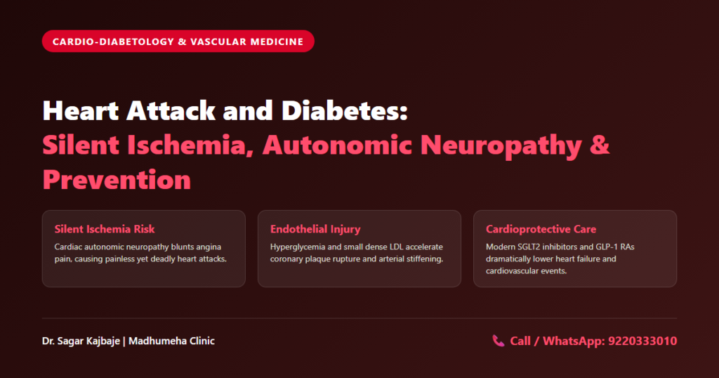
Heart Attack and Diabetes Symptoms: The Unseen Connection
Most people picture a heart attack as sudden chest pain radiating down the left arm. That dramatic depiction often misleads patients in daily clinical practice. Chronically high blood sugar alters vascular biology while dulling normal autonomic sensory perception. Vessels narrow silently. Blood slows. Tissues suffocate. Arteries fail. When blood glucose stays elevated for months, coronary arteries sustain progressive damage without triggering typical discomfort. Recognizing atypical heart attack and diabetes symptoms is therefore a matter of life and death.
Sugar damages endothelium. Cells die. Vessels stiffen. Circulating glucose molecules bind to structural proteins throughout vascular walls over extended timeframes. Endothelial cells progressively lose their biological capacity to synthesize nitric oxide gas, forcing muscular arterial walls to stiffen under unrelenting hydrostatic pressure. Microscopic mechanical tears repeatedly form along the fragile coronary intima, enabling atherogenic small dense LDL particles to easily infiltrate and accumulate within vulnerable subendothelial arterial layers. Plaques accumulate. Lumens shrink. Over time, these lipid deposits narrow arterial channels, severely restricting the flow of oxygenated blood to working heart muscle.
After 15 years treating diabetes at Madhumeha Clinic in Panchpakhadi, Thane West and Kalyan, I regularly evaluate patients who survived major cardiac events without experiencing chest pressure. Their presentations follow a familiar pattern. They dismiss sudden shortness of breath as simple fatigue. They mistake cold forehead perspiration for routine hot flashes or sudden drops in glucose. They write off upper abdominal discomfort as ordinary gastric acidity. That diagnostic delay destroys myocardium.
The Presentation That Defied the Textbooks: Mr Subhash Naik
Early on a rainy Tuesday morning, Mr Subhash Naik (name changed), a 58-year-old retired port trust officer from Panchpakhadi, Thane West, arrived at my clinic. He had lived with Type 2 diabetes for 12 years with inconsistent medical follow-up. He felt zero chest discomfort. Instead, he reported vague morning nausea, clammy forehead perspiration, and sudden breathlessness while climbing a single flight of stairs. He suspected gas trouble. My clinical examination pointed toward acute coronary ischemia.
We recorded a 12-lead electrocardiogram right in our treatment room without delay. The electrocardiogram tracing revealed pronounced ST-segment elevation across anterior precordial leads V2 through V4. An acute occlusive coronary thrombus was rapidly starving his anterior ventricular myocardium of oxygen, precipitating an evolving extensive anterior wall myocardial infarction with profound ischemic threat. Nerves lose feeling. Sensation ceases. Pain vanishes. Alarms stop. Cardiac autonomic neuropathy had blunted his sensory perception, producing a painless silent myocardial infarction.
Prompt action saves myocardium. Speed protects muscle. I initiated emergency dual antiplatelet therapy and statin loading before transferring him directly to the catheterization laboratory for emergency primary angioplasty. The interventional team reopened a 99 percent thrombotic occlusion within 40 minutes. He survived because we recognized atypical heart attack and diabetes symptoms rather than waiting for classic chest pain.
Unmasking Silent Heart Attack Diabetes: Why Nerve Damage Masks Pain
The Pathophysiology of Cardiac Autonomic Neuropathy (CAN)
A healthy heart produces intense ischemic pain when coronary blood flow halts. Oxygen-deprived cardiomyocytes switch to anaerobic glycolysis, releasing lactic acid, bradykinin, and adenosine into extracellular fluid. These chemical mediators stimulate visceral sensory afferent fibers traveling through cervical and upper thoracic sympathetic ganglia to the brainstem. The cerebral cortex interprets these intense impulses as crushing retrosternal pain. Angina pain disappears in long-standing diabetes.
Chronic hyperglycemia damages this neural alarm system through metabolic and microvascular injury. Excess intracellular glucose aggressively activates the aldose reductase polyol pathway, rapidly depleting indispensable cellular NADPH cofactors while generating a catastrophic cascade of reactive oxygen species. This relentless oxidative surge damages the vasa nervorum, the microscopic arterioles that nourish delicate autonomic visceral nerve trunks. Sensory vagal and sympathetic nerve fibers undergo relentless axonal degeneration and segmental demyelination, completely extinguishing visceral afferent impulses traveling toward the central nervous system.
This sensory blunting causes silent heart attack diabetes. When coronary blood supply fails, ischemic heart muscle releases pain-inducing chemicals, but the damaged nerve fibers cannot transmit the message. Myocardial necrosis and irreversible cardiomyocyte apoptosis progress silently beneath intact skin, entirely unhindered by the bodily warning alarms that normally drive emergency medical presentation. Clinical studies indicate that up to one in three diabetic patients suffers an acute myocardial infarction without noticeable angina.
Subtle Warning Signs: Recognizing Heart Attack and Diabetes Symptoms Early
Patients and family members must watch for subtle substitutes. Sudden shortness of breath during modest activity serves as the most frequent presentation. Unexplained cold sweats, sudden fatigue, throat tightness, or upper abdominal nausea must trigger prompt medical evaluation. Paying vigilant attention to subtle heart attack and diabetes symptoms prevents catastrophic treatment delays.
How Does Diabetes Cause Heart Disease and Endothelial Injury?
Accelerated Coronary Atherosclerosis and Plaque Rupture
Patients in my Thane clinic often ask: how does diabetes cause heart disease when diabetes is primarily a blood sugar issue? The answer resides in the vascular endothelium. The vascular endothelium is not an inert pipe lining. It functions as a metabolically active organ controlling arterial tone, platelet adhesion, and smooth muscle proliferation. Chronic hyperglycemia disrupts endothelial balance.
Intracellular glucose overload floods the mitochondrial electron transport chain with pyruvate, generating excessive superoxide free radicals. These free radicals degrade nitric oxide, which normally relaxes arteries. Endothelial nitric oxide synthase uncouples in the presence of intense oxidative stress, producing cytotoxic peroxynitrite free radicals rather than the physiological nitric oxide gas required for vasodilation. Vessels constrict. Pressure spikes. Vessels strain. Arterial stiffness accelerates systemic hypertension.
Circulating glucose also binds non-enzymatically to proteins and lipids, creating advanced glycation end-products. These harmful compounds cross-link structural collagen fibers within arterial walls, rendering coronary vessels rigid, fragile, and prone to injury. This process demonstrates how systemic inflammation accelerates cardiovascular damage across multiple vascular territories. Inflammatory signaling molecules draw circulating monocytes into the fragile subendothelial space of coronary arteries. These cells ingest oxidized lipids and transform into foam cells. Atherosclerotic plaques expand.
Atherogenic dyslipidemia drives this transformation further. Insulin resistance stimulates hepatic production of very low-density lipoproteins and triglycerides while reducing protective HDL particles. Circulating LDL particles become small and dense. Small dense LDL penetrates damaged endothelial layers with remarkable ease. Once inside the arterial wall, these lipids oxidize rapidly, generating unstable, necrotic plaque cores capped by thin fibrous tissue. Plaques rupture suddenly. Clots form. Flow stops. Muscle dies. Platelets aggregate instantly over the exposed core, forming an occlusive thrombus that abruptly halts coronary blood flow.
Diabetic Cardiomyopathy: The Non-Obstructive Danger to Heart Muscle
Understanding Heart Failure with Preserved Ejection Fraction (HFpEF)
Coronary artery blockages do not account for all cardiovascular complications in diabetes. High blood sugar damages myocardial tissue directly, even in the complete absence of epicardial coronary artery disease or hypertension. Physicians designate this condition as diabetic cardiomyopathy. The disorder alters myocardial metabolism and myocardial architecture, causing ventricular stiffness and pump dysfunction.
Under normal physiological conditions, healthy human myocardium demonstrates remarkable metabolic flexibility, constantly toggling between circulating blood glucose and plasma free fatty acids to generate adenosine triphosphate. In chronically diabetic metabolic states, severe myocardial insulin resistance forces cardiomyocytes to rely almost exclusively upon beta-oxidation of fatty acids, dramatically driving up myocardial oxygen consumption while generating toxic lipid intermediates that trigger lipotoxicity. Mitochondria lose efficiency. Energy drains. Intracellular calcium handling falters, preventing muscle fibers from relaxing properly between contractions.
Simultaneously, microvascular rarefaction decreases myocardial capillary density. Advanced glycation end-products cross-link delicate myocardial titin filaments, the microscopic biomechanical protein springs that determine ventricular diastolic compliance and physiological passive filling. Interstitial collagen deposition produces diffuse fibrosis throughout ventricular walls. The left ventricle becomes rigid. Although the heart muscle may contract with a normal ejection fraction, it cannot relax to fill with blood. This structural stiffening produces Heart Failure with Preserved Ejection Fraction, causing exertional breathlessness and fluid overload.
Unraveling Stiff Heart Syndrome: Mrs Pratibha Shinde
Mrs Pratibha Shinde (name changed), a 63-year-old homemaker from Kalyan, visited my clinic with progressive ankle edema, profound exhaustion, and orthopnea. She required three pillows to sleep comfortably without waking up breathless. She had managed Type 2 diabetes for 14 years, maintaining an average HbA1c above 8.8 percent. Her previous medical team performed an angiogram, suspecting major epicardial blockages. The angiogram revealed entirely patent coronary arteries.
Her breathlessness worsened steadily despite these reassuring coronary imaging findings. We conducted a detailed transthoracic echocardiogram with tissue Doppler imaging at our center. The scan confirmed a preserved ejection fraction of 54 percent. Tissue Doppler demonstrated severe diastolic impairment, elevated left ventricular filling pressures, and left atrial enlargement. Her serum N-terminal pro-B-type natriuretic peptide measured 1,480 pg/mL. Diabetic cardiomyopathy had stiffened her ventricle.
We restructured her medical regimen immediately. I initiated empagliflozin to promote decongestion and improve myocardial energetics. We added an angiotensin receptor-neprilysin inhibitor, refined her blood pressure therapy, and stabilized glucose levels. Within 14 weeks, her peripheral edema resolved completely. Her serum natriuretic peptide concentrations dropped by more than 60 percent over three months. She resumed her morning walks without shortness of breath.
Type 2 Diabetes and Cardiovascular Disease: The Risk Equivalent Reality
Why Diabetes Redefines Cardiac Risk Stratification
International cardiology societies classify type 2 diabetes and cardiovascular disease as clinical risk equivalents. This consensus means an adult with Type 2 diabetes carries the same statistical risk of suffering a heart attack as a patient with confirmed prior coronary disease. Diabetes is not merely an isolated metabolic condition. It represents a progressive vascular disease that compromises arterial function throughout the human body.
Persistent hyperglycemia establishes a severe prothrombotic hematological state, wherein hyperreactive platelets rapidly aggregate and adhere across vulnerable endothelial surfaces at minimal shear stress. Plasma fibrinogen concentrations rise, while endogenous fibrinolytic activity declines. Blood becomes more viscous. When a plaque ruptures inside a coronary vessel, these hematological changes accelerate complete thrombus formation. Ischemic damage escalates quickly. Time is muscle. Seconds count.
Routine cardiovascular screening remains necessary for every patient living with diabetes. At Madhumeha Clinic, we screen for early kidney microalbuminuria, which directly mirrors systemic endothelial injury. We evaluate C-reactive protein and review understanding your HbA1c test results and cardiovascular targets quarterly. We systematically screen for nocturnal respiratory abnormalities, recognizing how sleep apnea increases cardiovascular and metabolic strain while driving resistant nighttime hypertension in diabetic adults. Careful risk management saves lives. Vigilance protects. Testing saves. Maintaining blood pressure below 130/80 mmHg and lowering LDL cholesterol beneath 70 mg/dL stabilize fragile plaques. Recognizing subtle heart attack and diabetes symptoms provides a dependable final defense.
Cardioprotective Diabetology: Protecting the Heart Beyond Glucose Control
Modern Pharmacotherapy with SGLT2 Inhibitors and GLP-1 Receptor Agonists
Modern diabetology looks beyond glycemic lowering. Cardiovascular outcome trials over the past decade shifted our therapeutic paradigm toward organ-protective medicine. We now select antidiabetic medications for their proven power to prevent heart failure, halt atherosclerosis, and decrease mortality.
Sodium-glucose cotransporter-2 inhibitors provide profound cardiovascular risk reduction, with landmark multicenter randomized trials – such as EMPA-REG OUTCOME, DAPA-HF, and DECLARE-TIMI 58 – demonstrating sustained 30 to 35 percent reductions in heart failure hospitalizations across diverse patient populations. These agents induce mild osmotic diuresis and natriuresis, lowering ventricular preload and afterload. They shift myocardial substrate utilization toward efficient ketone bodies, generating cellular energy with reduced oxygen expenditure. Under this optimized energetic substrate paradigm, failing left ventricles pump with markedly heightened biomechanical efficiency while demanding far lower myocardial oxygen consumption.
Glucagon-like peptide-1 receptor agonists provide powerful anti-atherosclerotic vascular protection. Cardiovascular trials like LEADER and SUSTAIN-6 confirmed that GLP-1 receptor agonists reduce major adverse cardiovascular events by 12 to 26 percent. These cardioprotective agents directly restore endothelial integrity, quell systemic vascular inflammation, impede macrophage foam cell formation, and facilitate clinically meaningful, sustained body weight reduction. Selecting the appropriate agent depends on individual renal, cardiac, and metabolic profiles, which we explore when choosing between SGLT2 and GLP-1 medications.
Clinical Protocol Matrix: Cardiovascular Protection in Type 2 Diabetes
| Cardiovascular Disease Domain | Pathophysiological Mechanism & Patient Risk | Evidence-Based Medical Protocol | Target Cardio-Metabolic Outcome |
|---|---|---|---|
| Silent Myocardial Ischemia & Autonomic Neuropathy | Visceral sensory nerve demyelination blunts angina, delaying emergency presentation during acute coronary occlusions. | Baseline resting 12-lead ECG, annual cardiac autonomic testing, Holter monitoring for unexplained fatigue, and prompt coronary evaluation for atypical symptoms. | Early detection of silent ischemia, rapid emergency revascularization, and prevention of sudden ischemic mortality. |
| Accelerated Coronary Atherosclerosis & Endothelial Injury | Endothelial eNOS uncoupling, reactive oxygen species, and oxidized small dense LDL form rupture-prone coronary plaques. | Intensive statin therapy with ezetimibe, tight blood pressure control under 130/80 mmHg, antiplatelet therapy for secondary prevention, and HbA1c optimization. | Plaque stabilization, endothelial repair, and prevention of acute thrombotic coronary occlusion. |
| Diabetic Cardiomyopathy & Diastolic Dysfunction (HFpEF) | Microvascular rarefaction, myocardial titin glycation, and interstitial fibrosis create non-compliant ventricular walls independent of CAD. | Early SGLT2 inhibitor therapy, ARNI or ACEi/ARB administration, mineralocorticoid receptor antagonists, and metabolic decongestion. | Reduction in left ventricular filling pressures, reversal of diastolic stiffness, and prevention of heart failure hospitalizations. |
| Cardioprotective Pharmacotherapy (SGLT2i & GLP-1 RA) | Hemodynamic stress, renal sodium retention, systemic vascular inflammation, and progressive atherosclerotic burden. | Guideline-directed initiation of SGLT2 inhibitors (empagliflozin/dapagliflozin) and GLP-1 receptor agonists (semaglutide/liraglutide) alongside standard therapy. | 35% reduction in heart failure admissions, 12-26% reduction in 3-point MACE, and sustained renal preservation. |
Frequently Asked Questions About Heart Attack and Diabetes
1. Why are diabetics more likely to have silent heart attacks?
Patients with long-standing diabetes often develop cardiac autonomic neuropathy. Chronically high blood glucose damages the tiny blood vessels that nourish visceral sensory nerves traveling between the heart and the brain. This nerve demyelination prevents pain signals from registering in the cerebral cortex during an acute coronary occlusion. As a result, myocardial ischemia progresses without noticeable chest pressure, leading to unrecognized cardiac injury.
2. What are the subtle warning signs of heart attack in diabetic patients?
Diabetic patients frequently experience atypical symptoms instead of retrosternal pain. The most common manifestations include sudden breathlessness during light activity, cold forehead sweating, and unexplained nausea. Sudden profound exhaustion, inexplicable lightheadedness, or persistent dull aching across the jaw, throat, and interscapular region warrant urgent suspicion, demanding an immediate 12-lead electrocardiogram to rule out acute myocardial ischemia.
3. How does diabetes cause heart disease and arterial damage?
Diabetes damages arteries through persistent metabolic stress and endothelial injury. Elevated blood sugar generates reactive oxygen species that degrade nitric oxide, impairing healthy blood vessel dilation. Advanced glycation end-products cross-link structural collagen, making coronary vessels stiff and fragile. Additionally, insulin resistance creates atherogenic dyslipidemia with small dense LDL particles that penetrate arterial walls, oxidize, and build fragile, rupture-prone plaques.
4. Is diabetic cardiomyopathy reversible or preventable?
Diabetic cardiomyopathy can be prevented and partially reversed when managed early before dense myocardial fibrosis forms. Initiating SGLT2 inhibitors shifts cardiac metabolism toward efficient ketone bodies while unloading elevated ventricular filling pressures. Combining neurohormonal blockade with tight blood glucose control restores diastolic compliance. Once extensive collagen scarring replaces healthy myocytes, structural stiffness becomes permanent, making early screening necessary.
5. How do SGLT2 inhibitors and GLP-1 receptor agonists protect the heart in type 2 diabetes?
These two medication classes protect cardiovascular health through complementary biological pathways. SGLT2 inhibitors enhance urinary sodium and glucose excretion, reducing plasma volume, cardiac preload, and arterial afterload. They improve myocardial energetics and lower heart failure hospitalizations by up to 35 percent. GLP-1 receptor agonists act directly on blood vessel walls by decreasing vascular inflammation, stabilizing atherosclerotic plaques, and reducing major adverse cardiovascular events by up to 26 percent.
Patient Action Plan and Expert Diabetes Care
Living with diabetes requires a proactive approach to protect heart health before symptoms emerge. If you or a family member manages blood sugar, do not wait for chest pain to sound an alarm. Arrange regular cardiovascular assessments, review modern organ-protective medications with your physician, and treat sudden physical changes seriously.
Our clinical team provides thorough in-person evaluations at Madhumeha Clinic in Thane and Kalyan, along with accessible remote medical consultations. Patients across Maharashtra and India can schedule a specialized diabetes tele-consultation for personalized care. To book an appointment, please Call: 9220333010 or contact us via WhatsApp: 9220333010. You can also view Madhumeha Clinic on Google Maps for route guidance to our Panchpakhadi location.
Consulting Diabetologist & Clinical Lead:
Dr Sagar Kajbaje
DNB (General Medicine), Post Graduate Diploma in Diabetes and Endocrinology, Royal College of Physicians (UK)
Practicing Senior Diabetologist with 15+ years of clinical experience in Thane and Kalyan, Maharashtra.
[…] Coronary Artery Disease & Heart Attack: Visceral fat promotes arterial plaque rupture and silent ischemia. Understand the shared vascular mechanisms in our clinical analysis on the link between heart attack and diabetes. […]
[…] spikes strain fragile cardiac vessels. Sudden wall tension triggers plaque rupture, creating an increased vulnerability to cardiovascular events and heart attack during early waking hours. Myocardial oxygen demand jumps while coronaries struggle to supply […]